Back Of Skull Anatomy Labeled - Skull Anatomy / Skull, skeletal framework of the head of vertebrates, composed of bones or cartilage, which form a unit that protects the brain and some sense organs.
Back Of Skull Anatomy Labeled - Skull Anatomy / Skull, skeletal framework of the head of vertebrates, composed of bones or cartilage, which form a unit that protects the brain and some sense organs.. Please feel free to download and print. Examine the cranial bones of the articulated human skull and the sectioned skull. Skull reshaping is done on any of the structures that lie above the face. Start studying anatomy skull labels. 12 photos of the bone of back of skull.
Overview, anterior skull base, middle skull base march 18, 2017. The skull performs vital functions. The skull is the bony skeleton of the head. If you'd like to customize what appears on the front and back of a card, you. Adelstein on skull labeling anatomy:
The Skull Anatomy Physiology from pressbooks-dev.oer.hawaii.edu Please feel free to download and print. Learn vocabulary, terms and more with flashcards, games and other study tools. It supports and protects the face and the brain. Anatomy and physiology7.2 the skull. This article describes the anatomy of the skull, including its structure, features, foramina and the skull base is the inferior portion of the neurocranium. It offers protection to the brain, eye balls, inner ears, and nasal passages. The skull includes the upper jaw and the cranium. Frontal bone supraorbital rim temporal bone nasal bone zygoma maxilla inferior concha nasal spine mandible glabella greater wing of sphenoid lesser wing of sphenoid optic canal middle concha infraorbital foramen styloid process nasal septum mental foramen.
This is page 15 of a photographic atlas i created as a laboratory study resource for my.
The skull or known as the cranium in the medical world is a bone structure of the head. When this deck is imported into the desktop program, cards will appear as the deck author has made them. Anatomy visible in the medical illustration includes: That is how the doctor insights on: Exterior skull anatomy 3d model. Anatomical structures of the skull include: This is a model of the human (homo sapiens) skull. Learn vocabulary, terms and more with flashcards, games and other study tools. The skull includes the upper jaw and the cranium. Adelstein on skull labeling anatomy: 3d viewer is not available. We also cover the ear bones and the hyoid bone.transcript/notesskull. The skull performs vital functions.
Anatomy visible in the medical illustration includes: This is a model of the human (homo sapiens) skull. Learn more here you are seeing a 360° image instead. Learn more about the anatomy and function of the skull in humans and other vertebrates. It was then cleaned, adapted and polypainted this model is part of a comparison with the skull of a human.
Skull Functions Facts Fractures Protection View Bones from i1.wp.com It supports and protects the face and the brain. Anatomy visible in the medical illustration includes: Related posts of bone of back of skull. Excluding ear ossicles, it is made of 22 bones. Learn vocabulary, terms and more with flashcards, games and other study tools. It is comprised of many bones, formed by intramembranous ossification, which are joined together by sutures (fibrous joints). As a review activity, label figures 13.1, 13.2, 13 3, 13.4, and 13.5. That is how the doctor insights on:
Skull reshaping is done on any of the structures that lie above the face.
3d viewer is not available. We also cover the ear bones and the hyoid bone.transcript/notesskull. Start studying anatomy skull labels. As a review activity, label figures 13.1, 13.2, 13 3, 13.4, and 13.5. Exterior skull anatomy 3d model. Human skull anatomy, the skeletal structure of the highest point of vertebrates, made out of bones or ligament, which shapes a unit that, ensures the brain and some vibe organs. Adelstein on skull labeling anatomy: Learn skull anatomy with skull bones quizzes and diagram labeling exercises. The skull is the bony skeleton of the head. At the same time the bones grow larger by growing back into the growth plates. The frontal, parietal, temporal and occipital bones are joined at the cranial sutures. Anatomy and physiology7.2 the skull. Looking at it from the inside it can be learn everything about the bones of the skull with our articles, video tutorials, labeled diagrams, and quizzes.
Learn about the anatomy of the skull bones and sutures as seen on ct images of the brain. Anatomy and physiology7.2 the skull. We also cover the ear bones and the hyoid bone.transcript/notesskull. In order to be light, the skull is made up by flat and irregular bones, and has hollow spaces called the sinuses. It was then cleaned, adapted and polypainted this model is part of a comparison with the skull of a human.
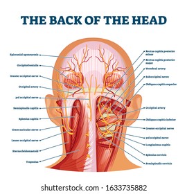
Occipital Hd Stock Images Shutterstock from image.shutterstock.com We use cookies to ensure that we give you the best experience on our website. This anatomic region is complex and poses surgical challenges for otolaryngologists and neurosurgeons alike. The major sutures are the coronal suture, sagittal suture, lambdoid suture and squamosal sutures. If you'd like to customize what appears on the front and back of a card, you. Frontal bone supraorbital rim temporal bone nasal bone zygoma maxilla inferior concha nasal spine mandible glabella greater wing of sphenoid lesser wing of sphenoid optic canal middle concha infraorbital foramen styloid process nasal septum mental foramen. The human head, the component that incorporates the. Anatomy visible in the medical illustration includes: 12 photos of the bone of back of skull.
All the bones of skull, joined together by sutures… the skull is subdivided into 2 parts:
The skull is the bony skeleton of the head. The skull is a bony structure that supports the face and forms a protective cavity for the brain. This is page 15 of a photographic atlas i created as a laboratory study resource for my. Skull, skeletal framework of the head of vertebrates, composed of bones or cartilage, which form a unit that protects the brain and some sense organs. They don't move and united into a single unit. Exterior skull anatomy 3d model. In order to be light, the skull is made up by flat and irregular bones, and has hollow spaces called the sinuses. Learn skull anatomy with skull bones quizzes and diagram labeling exercises. Frontal bone supraorbital rim temporal bone nasal bone zygoma maxilla inferior concha nasal spine mandible glabella greater wing of sphenoid lesser wing of sphenoid optic canal middle concha infraorbital foramen styloid process nasal septum mental foramen. Please feel free to download and print. Anatomy and physiology7.2 the skull. Skull, parietal bone, nasion, frontal process, lacrimal bone, nasal bone, spehenoid lesser wing, middle nasal. Examine the cranial bones of the articulated human skull and the sectioned skull.


0 Komentar